II · THE DISCOVERY · HISTORY OF SCIENCE
Red blood cell
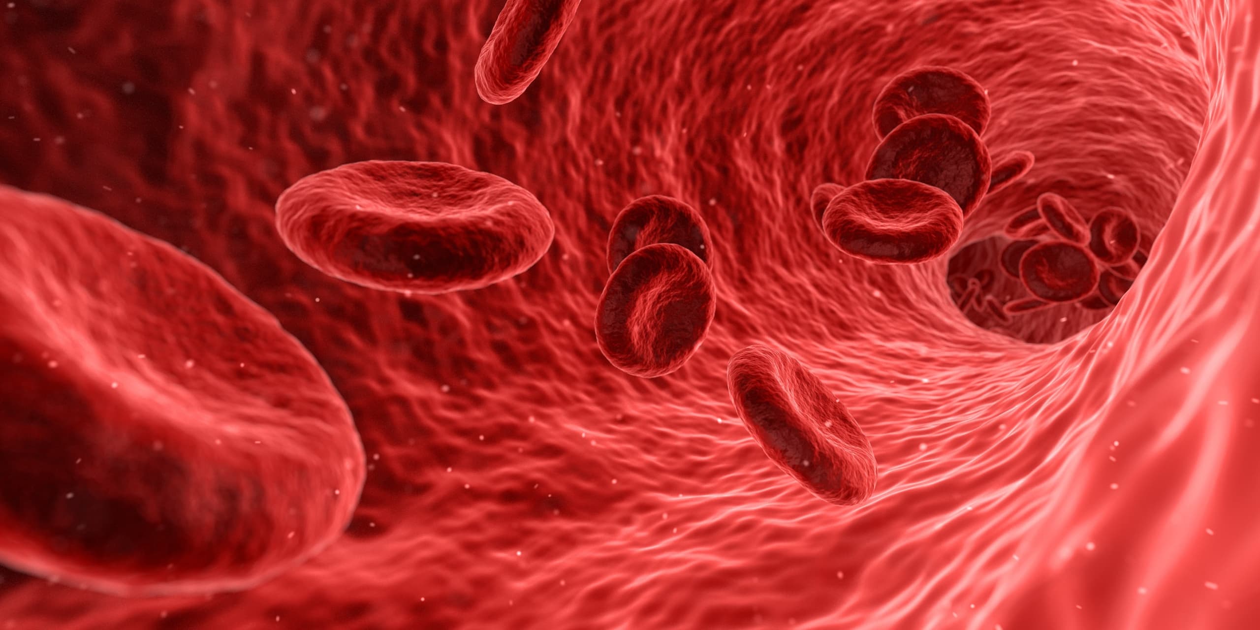
The cells are seven micrometres across — small enough that you need a microscope to see one, numerous enough that nearly half the volume of your blood is made of them.
At a glance
- Discovered
- 1658
- Discoverer
- Jan Swammerdam
- Diameter
- 7 micrometres
- Type
- Cell type
Look closer
The size is a constraint
Seven micrometres is small enough to pass through the narrowest vessels in the body — the capillaries, where oxygen leaves the blood and enters the tissues. The cell is flexible as well as small: it deforms as it squeezes through, then returns to shape. That flexibility depends on the membrane, which is made of proteins and lipids arranged in a structure that bends without tearing.
The shape is biconcave
In humans, a red blood cell is a disc that curves inward on both faces, like a deflated ball pressed between two thumbs. The shape increases the surface area relative to the volume, which matters because oxygen has to cross the membrane to enter or leave. The cell has no nucleus and no internal structures — those are expelled during development — so the interior is nearly all hemoglobin, the molecule that binds oxygen.
The colour comes from iron
Hemoglobin is the protein that gives red blood cells their colour. Each molecule contains iron, and it is the iron that binds oxygen in the lungs and releases it in the tissues. Each red blood cell contains approximately 270 million hemoglobin molecules. The red colour is the iron's; the cell itself is essentially a membrane sack built to carry as much of it as possible.
The story
Red blood cells are the most common type of cell in vertebrate blood. They carry oxygen from the lungs — or, in fish, the gills — to the tissues, and they do it by squeezing through vessels so narrow that the cells deform as they pass.
Jan Swammerdam saw them in 1658. What he saw would have been the cells themselves, seven micrometres across, visible under a microscope but not to the unaided eye. What he could not have seen was their function: that required an understanding of oxygen, of circulation, and of the role of blood in moving gases through the body, none of which was settled in the mid-seventeenth century.
The cell is built around a single task. It has no nucleus — that is expelled during development — and no internal structures that other cells use to maintain themselves. What it has instead is hemoglobin, an iron-containing protein that binds oxygen. Each cell contains approximately 270 million hemoglobin molecules, packed into a flexible membrane that bends as the cell moves through capillaries and returns to shape afterward.
The shape is biconcave: a disc that curves inward on both faces. That shape increases the surface area relative to the volume, which matters because oxygen crosses the membrane by diffusion. The more surface, the faster the exchange. The cell picks up oxygen in the lungs, where the concentration is high, and releases it in the tissues, where the concentration is low. Each circuit through the body takes about sixty seconds.
In human adults, approximately 2.4 million new red blood cells are produced every second. They develop in the bone marrow, circulate for about one hundred to one hundred and twenty days, and are then broken down by macrophages — cells that consume and recycle cellular debris. The components are reused. Nearly half the volume of blood is red blood cells: between forty and forty-five per cent, depending on the individual.
The cells are also called erythrocytes, from the Greek for red and hollow vessel, though the vessel is not hollow in the sense of being empty. It is full of hemoglobin. The term is used in medical and academic writing; red blood cell is the plain version, and the one more common outside those contexts.
Why it mattered then
Swammerdam's observation in 1658 was the beginning of seeing blood as something with structure rather than as a uniform fluid. He saw the cells, but the explanation for what they did — carrying oxygen, binding it with iron, releasing it in tissues — came much later, as the chemistry of gases and the mechanics of circulation were worked out. The discovery mattered because it made blood a subject that could be studied at the level of its components, not just as a substance that flowed. What physicians could not do before 1658 was see that blood contained distinct, repeating units. Afterward, those units became something that could be counted, measured, and eventually understood as the means by which oxygen reaches the body's tissues.
Why it matters now
Red blood cells are central to transfusion medicine. Donated blood is separated into components, and packed red blood cells — the cells with most of the plasma removed — are stored in blood banks and used to treat patients who have lost blood or whose bodies do not produce enough cells of their own. The cells are also studied in disorders where their shape, number, or hemoglobin content is wrong: sickle cell disease, anaemias, and conditions where the bone marrow does not produce cells at the rate the body needs. Understanding the cell's structure — the membrane, the lack of a nucleus, the hemoglobin content — is necessary for diagnosing and treating those conditions. The discovery Swammerdam made in 1658 is the foundation for all of that work, though the path from seeing the cells to using them in medicine took centuries.
The surprising detail
The cells have no nucleus and no internal machinery for maintaining themselves, which means they cannot repair damage or reproduce. They are produced in the bone marrow, released into circulation, and broken down after about one hundred to one hundred and twenty days. In that sense, they are disposable: the body makes them, uses them, and recycles them continuously. Approximately 2.4 million are produced every second in a human adult, which is necessary because the same number are being destroyed. The cell is built to do one thing — carry oxygen — and it does it by being as simple as a cell can be while still functioning.
What is disputed
The verified facts give 1658 as the discovery date and Jan Swammerdam as the discoverer, but the reference material does not describe what Swammerdam saw or what instruments he used, so the lesson cannot state whether he observed the cells in human blood, animal blood, or under what magnification. The diameter of seven micrometres is recorded in Wikidata, but the reference material does not say whether that is a human red blood cell specifically or an average across species, so the lesson treats it as the human measurement where context requires it and leaves it unqualified elsewhere.
Remember this
Swammerdam saw the cells in 1658. What they did — carrying oxygen — took longer to work out.
Test yourself
Red blood cells have no nucleus and no internal structures. Why does that matter for what the cell does?
The lack of a nucleus and organelles means the interior of the cell is almost entirely hemoglobin — the protein that binds oxygen. The cell is essentially a membrane sack built to carry as much hemoglobin as possible, which maximises the amount of oxygen it can transport. The trade-off is that the cell cannot repair itself or reproduce, because it has none of the machinery other cells use for those tasks. It is produced in the bone marrow, circulates for about one hundred to one hundred and twenty days, and is then broken down and recycled. The simplicity is the design: the cell does one thing, and it is built to do that thing as efficiently as possible.